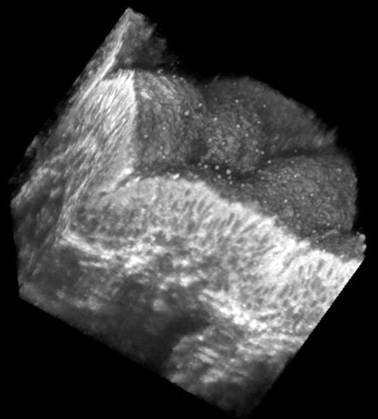
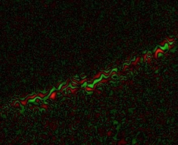
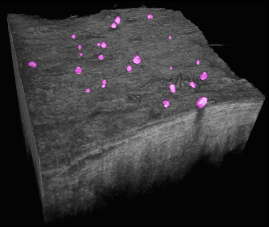
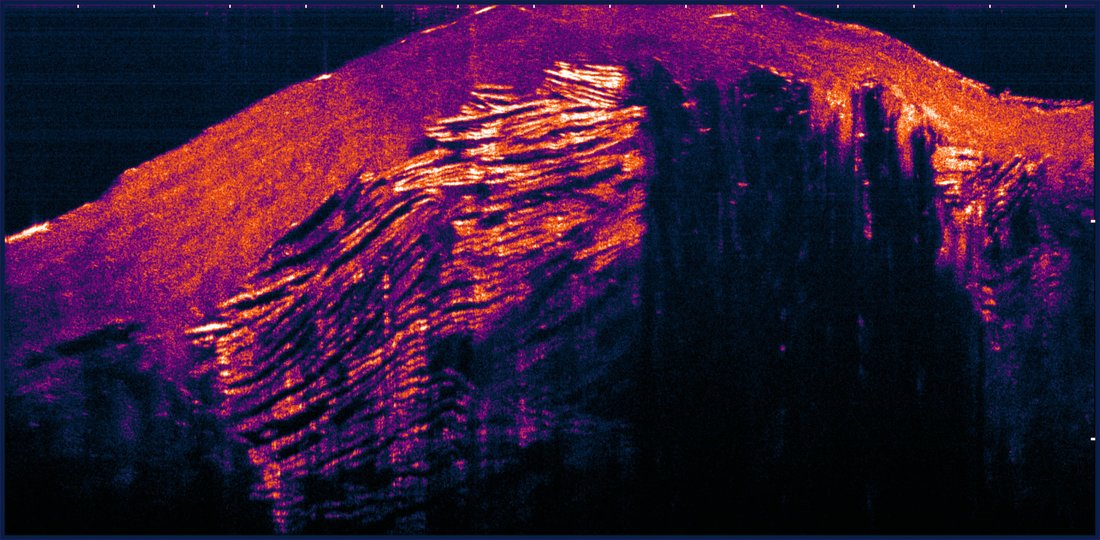
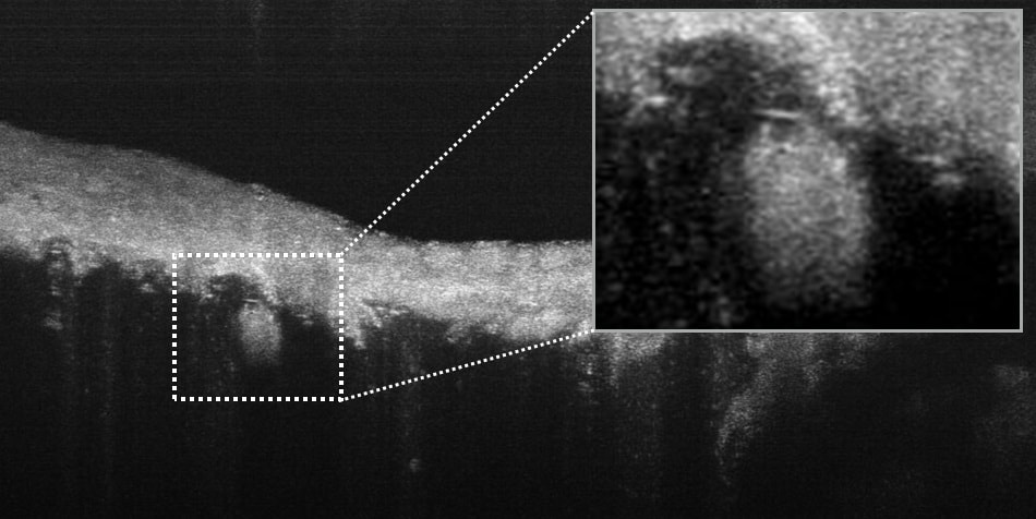
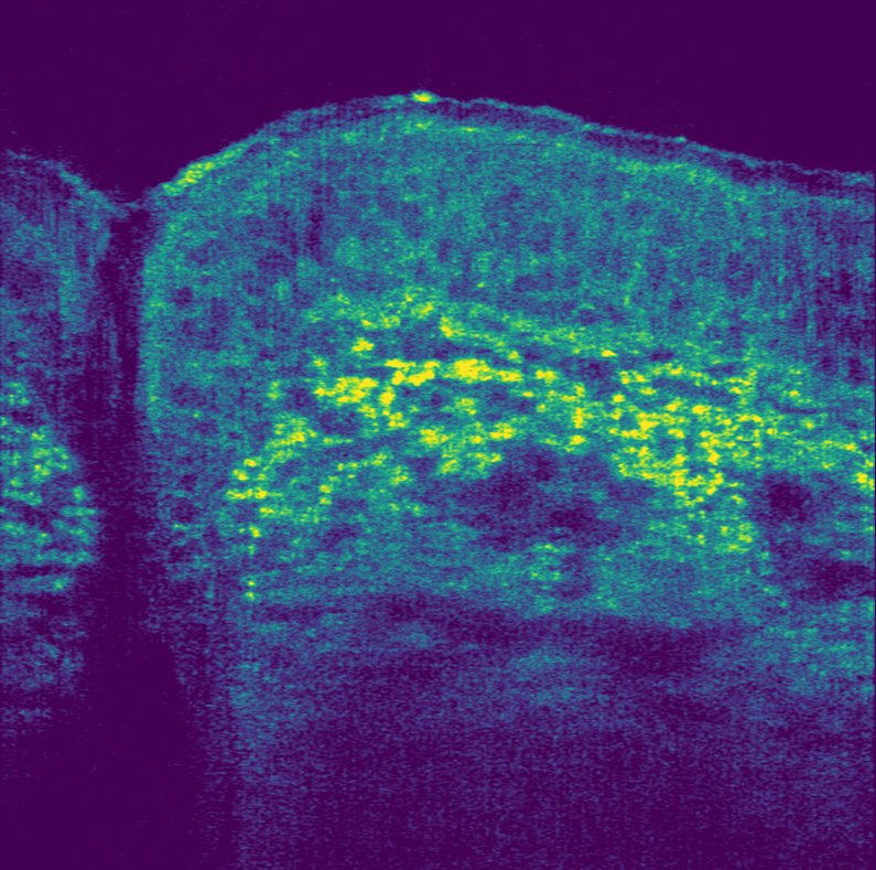
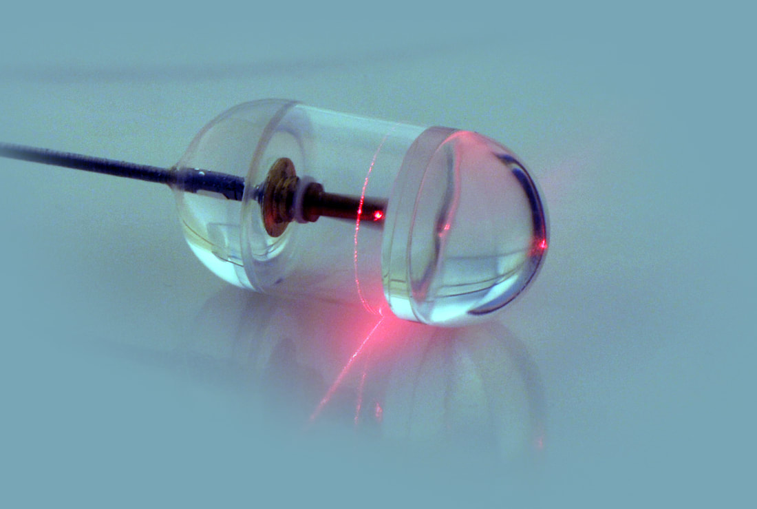
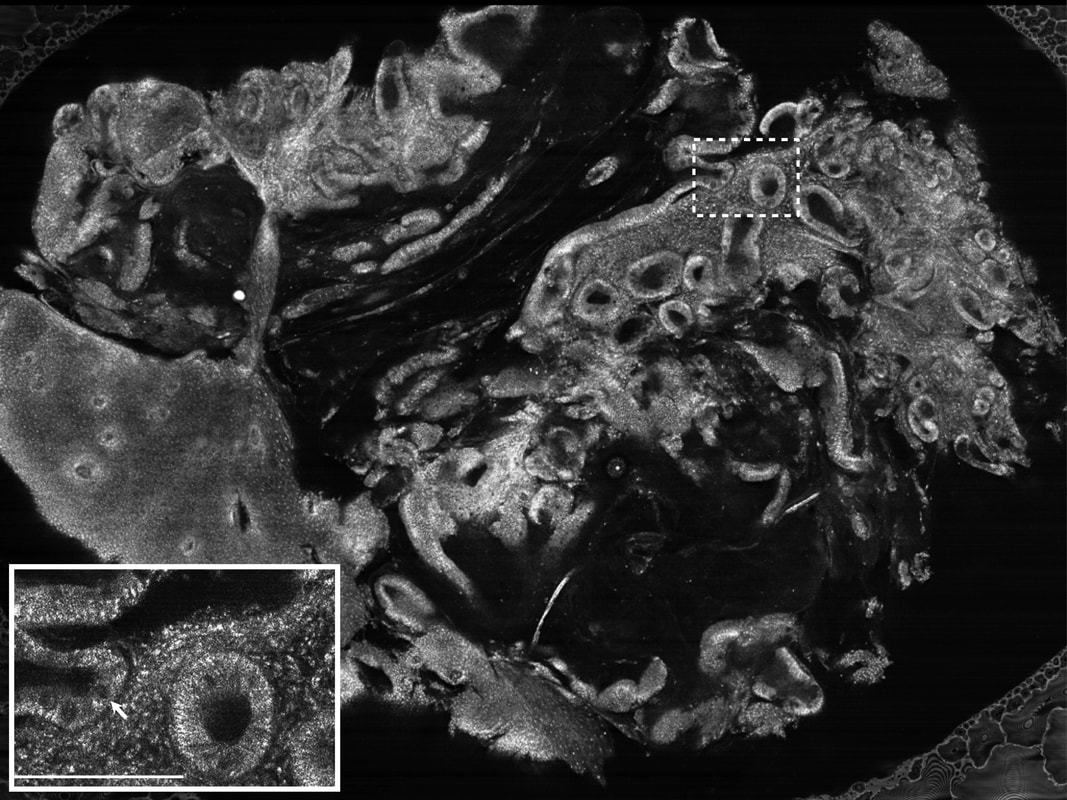
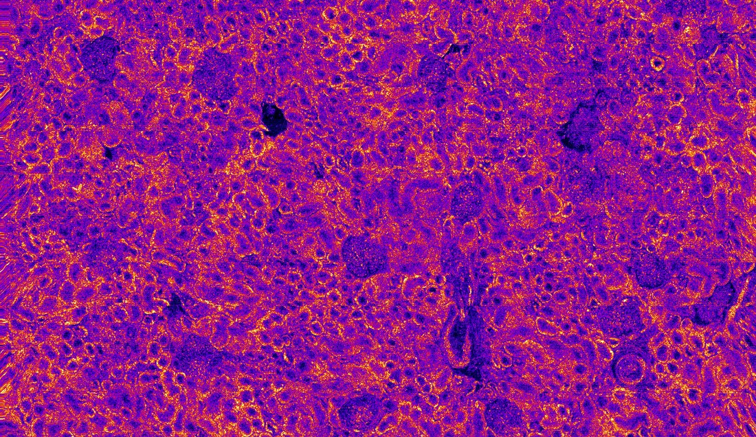
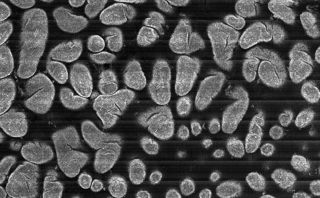
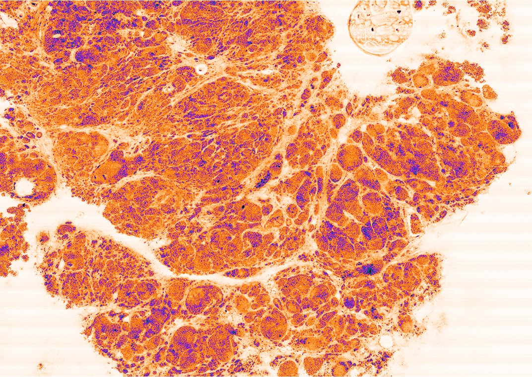
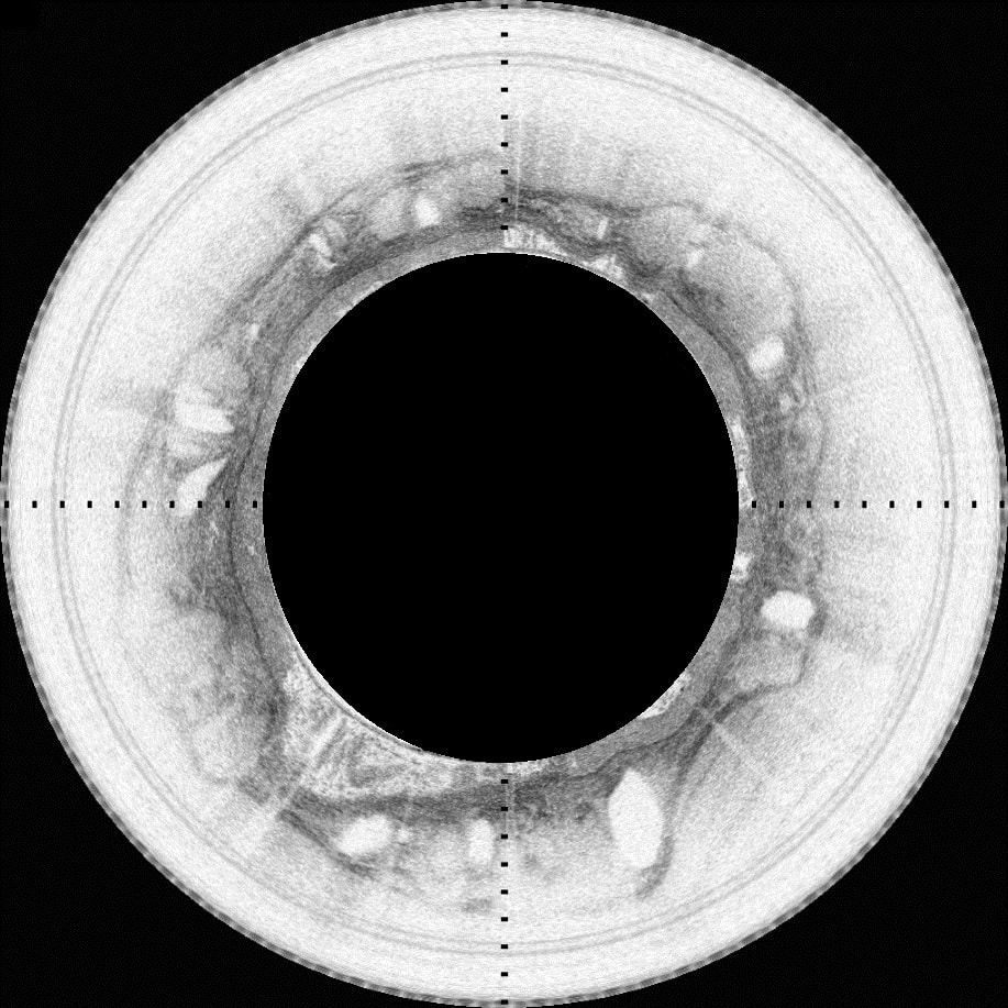
Micro-Optical Coherence Tomography (μOCT)
Micro-Optical Coherence Tomography (μOCT) is the highest resolution cross-sectional OCT technology available today. The high resolution and imaging speed of μOCT make it uniquely suited for imaging many biological phenomena. One such example is the respiratory airways that are lined with microscopic hairs called cilia that continuously work to sweep away mucus. This process is a crucial element of respiratory defense that becomes impaired in a variety of diseases, including Cystic Fibrosis. Another example is vascular disease where μOCT enables visualization of individual cells and their interactions, helping to discover the factors that contribute to coronary plaque formation, progression, and disruption.

Publications
Biwei Yin, Chulho Hyun, Joseph A. Gardecki, and Guillermo J. Tearney, Extended depth of focus for coherence-based cellular imaging, Optica 4, 959-965 (2017)
Chu KK, Kusek ME, Liu L, Som A, Yonker LM, Leung H, Cui D, Ryu J, Eaton AD, Tearney GJ, Hurley BP. Illuminating dynamic neutrophil trans-epithelial migration with micro-optical coherence tomography. Sci Rep. 2017 Apr 3;8:45789. doi: 10.1038/srep45789. PubMed PMID: 28368012; PubMed Central PMCID: PMC5377939.
Chu KK, Mojahed D, Fernandez CM, Li Y, Liu L, Wilsterman EJ, Diephuis B, Birket SE, Bowers H, Martin Solomon G, Schuster BS, Hanes J, Rowe SM, Tearney GJ. Particle-Tracking Microrheology Using Micro-Optical Coherence Tomography. Biophys 2016 Sep 6;111(5):1053-63. doi: 10.1016/j.bpj.2016.07.020. PubMed PMID: 27602733; PubMed Central PMCID: PMC5018123.
Biwei Yin, Kengyeh K. Chu, Chia-Pin Liang, Kanwarpal Singh, Rohith Reddy, and Guillermo J. Tearney. μOCT imaging using depth of focus extension by self-imaging wavefront division in a common-path fiber optic probe. Optics express 24, no. 5 (2016): 5555-5564.
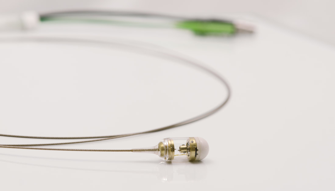
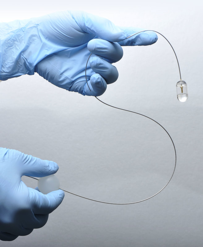
Capsule Technology
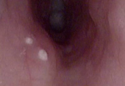
The Tearney Lab has unlocked a new paradigm for diagnosing gastrointestinal (GI) diseases that eliminates the sampling errors associated with endoscopic biopsy – all in a simple, rapid, and inexpensive procedure. The technique, termed tethered capsule endomicroscopy (TCE), involves swallowing an optomechanically-engineered pill that obtains microscopic images of the GI tract. The results obtained from unsedated human subjects showed that three-dimensional microscopic images of the entire esophagus can be obtained in just a few minutes. After imaging, the capsule is withdrawn using the tether, disinfected, and reused. The capsule is now being tested in the primary care setting to see if it can be utilized for Barrett’s Esophagus screening. Other applications being investigated include the diagnosis of Celiac Disease, Eosinophilic Esophagitis, and Environmental Enteropathy.

Publications
Tabatabaei N, Kang D, Kim M, Wu T, Grant CN, Rosenberg M, Nishioka NS, Hesterberg PE , Garber J, Yuan Q, Katz AJ, Tearney GJ. Clinical Translation of Tethered Confocal Microscopy Capsule for Unsedated Diagnosis of Eosinophilic Esophagitis. Sci Rep. 2018;8:2631
Gora MJ, Simmons LH, Quénéhervé L, Grant CN, Carruth RW, Lu W, Tiernan A, Dong J, Walker-Corkery B, Soomro A, Rosenberg M, Metlay JP, Tearney GJ. Tethered capsule endomicroscopy: from bench to bedside at a primary care practice. J Biomed Opt. 2016 Oct 1;21(10):104001. doi: 10.1117/1.JBO.21.10.104001. PubMed PMID: 27689919; PubMed Central PMCID: PMC5043371.
Gora MJ, Sauk JS, Carruth RW, Gallagher KA, Suter MJ, Nishioka NS, Kava LE, Rosenberg M, Bouma BE, Tearney GJ. Tethered capsule endomicroscopy enables less invasive imaging of gastrointestinal tract microstructure. Nat Med. 2013 Feb;19(2):238-40. doi: 10.1038/nm.3052. Epub 2013 Jan 13. PubMed PMID: 23314056; PubMed Central PMCID: PMC3567218.
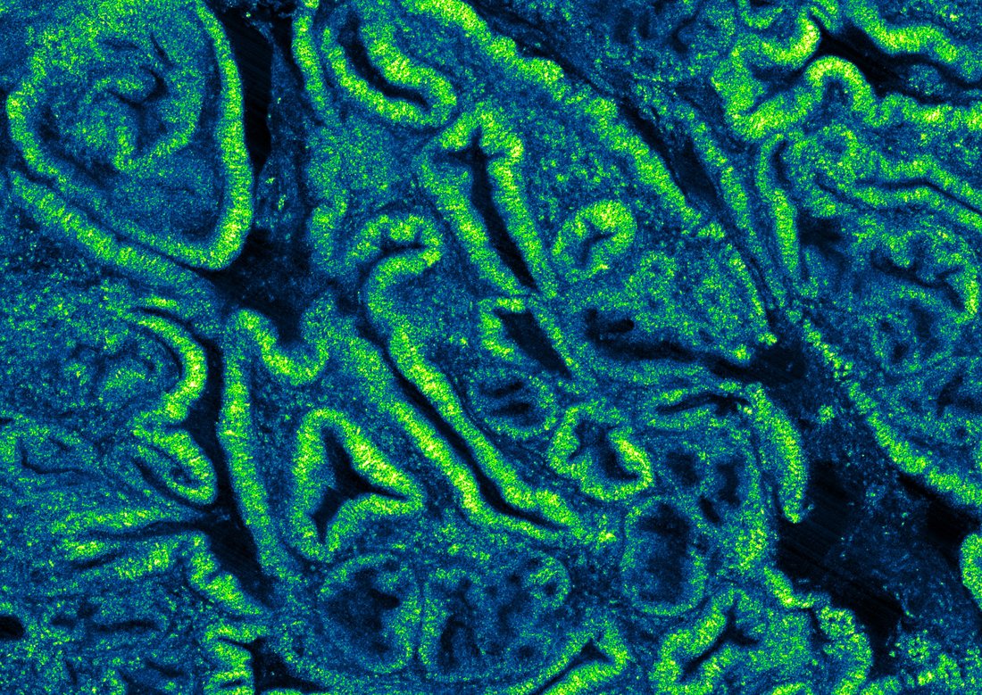
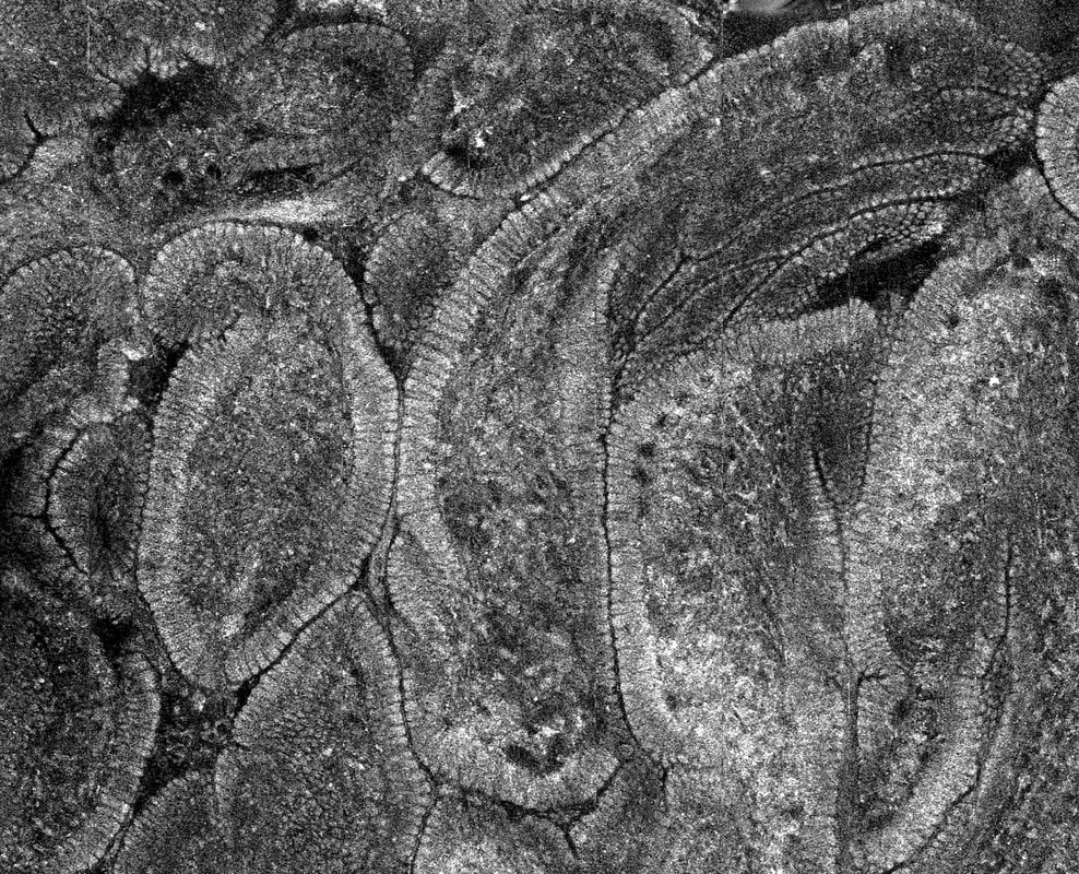
Spectrally Encoded Confocal Microscopy (SECM)
The Tearney laboratory has developed a new form of confocal microscopy, termed spectrally encoded confocal microscopy (SECM), that does not require integrated high-speed mechanical components, yet is capable of obtaining cellular-level resolution images at thousands of frames per second through an endoscope. The SECM team is fabricating endoscopic probes and capsules capable of imaging entire luminal organs with this technology.
Publications
Tabatabaei N, Kang D, Kim M, Wu T, Grant CN, Rosenberg M, Nishioka NS, Hesterberg PE , Garber J, Yuan Q, Katz AJ, Tearney GJ. Clinical Translation of Tethered Confocal Microscopy Capsule for Unsedated Diagnosis of Eosinophilic Esophagitis. Sci Rep. 2018;8:2631
Kang D, Schlachter SC, Carruth RW, Kim M, Wu T, Tabatabaei N, Soomro AR, Grant CN, Rosenberg M, Nishioka NS, Tearney GJ. Large-area spectrally encoded confocal endomicroscopy of the human esophagus in vivo. Lasers Surg Med. 2017 Mar;49(3):233-239. doi: 10.1002/lsm.22585. Epub 2016 Sep 16. PubMed PMID: 27636715.
Kang D, Schlachter SC, Carruth RW, Kim M, Wu T, Tabatabaei N, Soomro AR, Grant CN, Rosenberg M, Nishioka NS, Tearney GJ. Large-area spectrally encoded confocal endomicroscopy of the human esophagus in vivo. Lasers Surg Med. 2017 Mar;49(3):233-239. doi: 10.1002/lsm.22585. Epub 2016 Sep 16. PubMed PMID: 27636715.
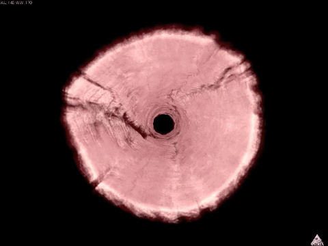
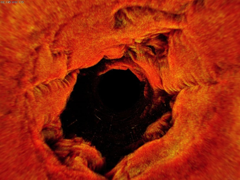
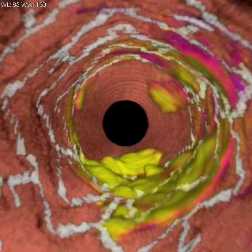
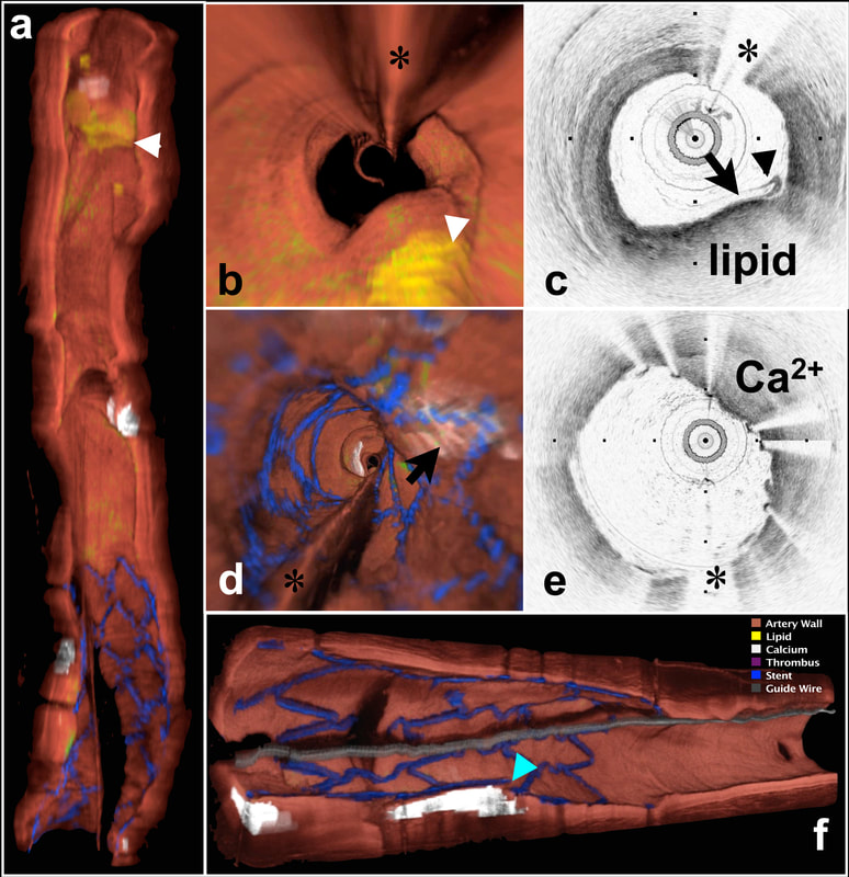
Optical Frequency-Domain Imaging (OFDI)
An advanced form of OCT called optical frequency-domain imaging (OFDI), also known as swept-source OCT (SS-OCT), achieves more than 50-fold improvement in image acquisition speed, compared to its preceding technology, time-domain OCT (TD-OCT). Our work with OFDI is focused on solving clinical dilemmas surrounding early detection of atherosclerosis and cancer.


Publications
Gora MJ, Sauk JS, Carruth RW, Gallagher KA, Suter MJ, Nishioka NS, Kava LE, Rosenberg M, Bouma BE, Tearney GJ. Tethered capsule endomicroscopy enables less invasive imaging of gastrointestinal tract microstructure. Nat Med. 2013 Feb;19(2):238-40. doi: 10.1038/nm.3052. Epub 2013 Jan 13. PubMed PMID: 23314056; PubMed Central PMCID: PMC3567218.
Suter MJ, Vakoc BJ, Yachimski PS, Shishkov M, Lauwers GY, Mino-Kenudson M, Bouma BE, Nishioka NS, Tearney GJ. Comprehensive microscopy of the esophagus in human patients with optical frequency domain imaging. Gastrointest Endosc. 2008 Oct;68(4):745-53. doi: 10.1016/j.gie.2008.05.014. PubMed PMID: 18926183; PubMed Central PMCID: PMC2715833.
Tearney GJ, Waxman S, Shishkov M, Vakoc BJ, Suter MJ, Freilich MI, Desjardins AE, Oh WY, Bartlett LA, Rosenberg M, Bouma BE. Three-dimensional coronary artery microscopy by intracoronary optical frequency domain imaging. JACC Cardiovasc Imaging. 2008 Nov;1(6):752-61. doi: 10.1016/j.jcmg.2008.06.007. PubMed PMID: 19356512; PubMed Central PMCID: PMC2852244.
