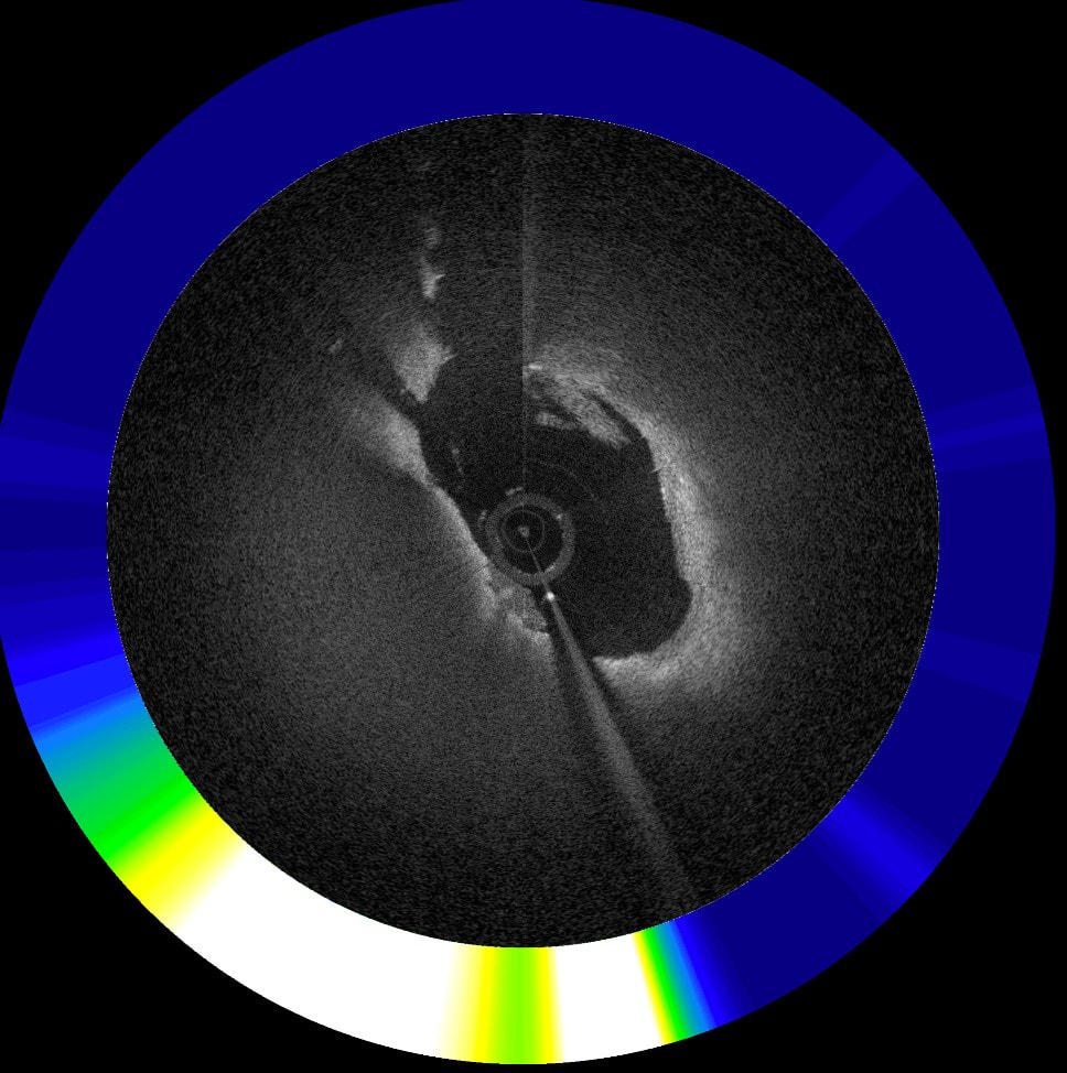
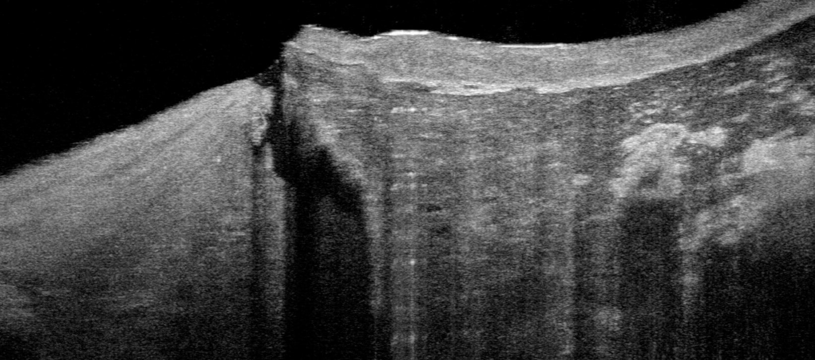
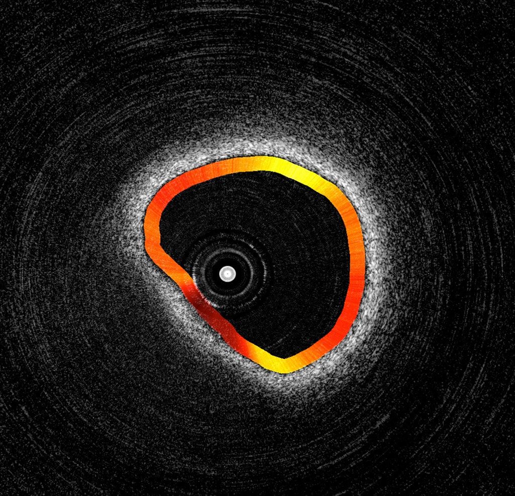
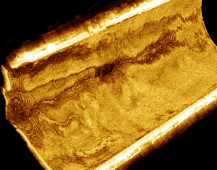
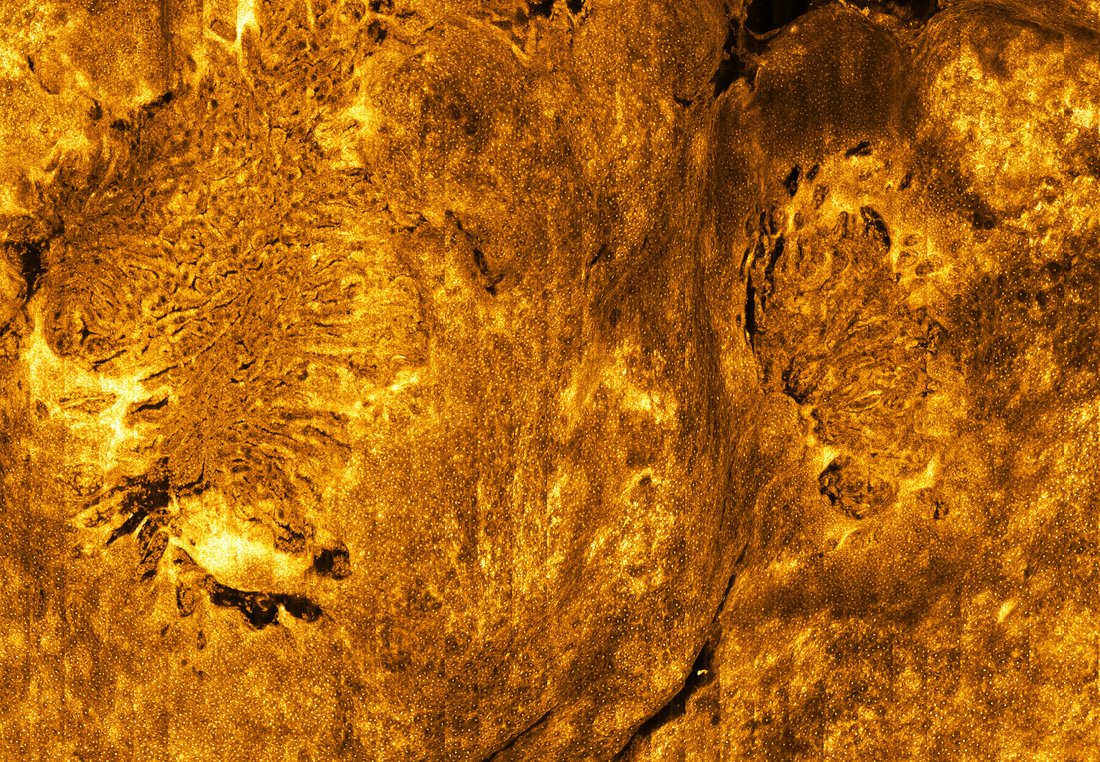
Coronary Artery Disease
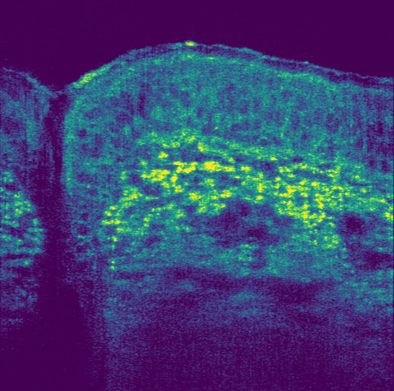
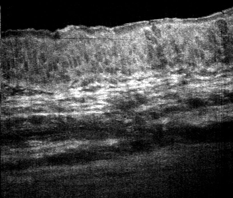
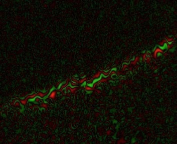
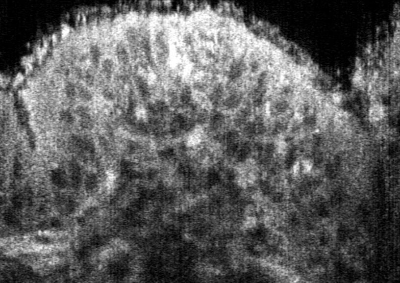
The importance of understanding coronary artery disease cannot be overstated; it is the number one cause of death in the US. Each year half a million Americans die from heart attacks. An equal number survive with considerable and permanent damage to their heart. Coronary artery disease is caused by cells that are too small to visualize using modern medical imaging technologies. We have developed multiple new technologies that enable the visualization of structural details, fluorescent and absorbing molecules, and individual cells in the coronaries of living patients. Our lab’s work with with advanced coronary imaging will change how we study, diagnose, and treat atherosclerosis, allowing clinicians to intervene before a heart attack occurs.


Publications
Ughi GJ, Wang H, Gerbaud E, Gardecki JA, Fard AM, Hamidi E, Vacas-Jacques P, Rosenberg M, Jaffer FA, Tearney GJ. Clinical Characterization of Coronary Atherosclerosis With Dual-Modality OCT and Near-Infrared Autofluorescence Imaging. JACC Cardiovasc Imaging. 2016 Nov;9(11):1304-1314. doi: 10.1016/j.jcmg.2015.11.020. Epub 2016 Mar 9. PubMed PMID: 26971006; PubMed Central PMCID: PMC5010789.
Fard AM, Vacas-Jacques P, Hamidi E, Wang H, Carruth RW, Gardecki JA, Tearney GJ. Optical coherence tomography--near infrared spectroscopy system and catheter for intravascular imaging. Opt Express. 2013 Dec 16;21(25):30849-58. doi: 10.1364/OE.21.030849. PubMed PMID: 24514658; PubMed Central PMCID: PMC3926541.
Liu L, Gardecki JA, Nadkarni SK, Toussaint JD, Yagi Y, Bouma BE, Tearney GJ. Imaging the subcellular structure of human coronary atherosclerosis using micro-optical coherence tomography. Nat Med. 2011 Jul 10;17(8):1010-4. doi:10.1038/nm.2409. PubMed PMID: 21743452; PubMed Central PMCID: PMC3151347.
Tearney GJ, Waxman S, Shishkov M, Vakoc BJ, Suter MJ, Freilich MI, Desjardins AE, Oh WY, Bartlett LA, Rosenberg M, Bouma BE. Three-dimensional coronary artery microscopy by intracoronary optical frequency domain imaging. JACC Cardiovasc Imaging. 2008 Nov;1(6):752-61. doi: 10.1016/j.jcmg.2008.06.007. PubMed PMID: 19356512; PubMed Central PMCID: PMC2852244.
Eosinophilic Esophagitis
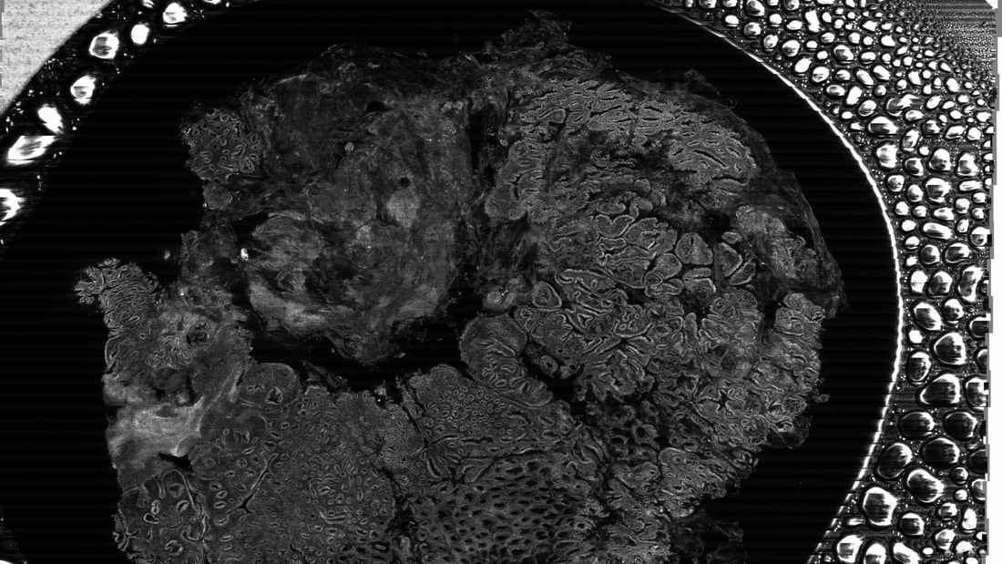
Eosinophilic esophagitis (EoE) is a common food allergy that is characterized by infiltration of eosinophils in the esophagus. EoE causes in symptoms such as nausea, vomiting, heartburn, food impactions, and difficulty swallowing. Current standard of care can require many sedated endoscopies with biopsies to diagnose and confirm elimination of the eosinophils following therapy. These procedures are expensive, time consuming, and can be difficult for patients to tolerate. We have recently developed a swallowable SECM capsule that is capable of identifying individual eosinophils in the esophagus without requiring sedation or use of contrast agents. This device has the potential to be a reliable and affordable tool for the diagnosis and follow up care of EoE patients.
Publications
Tabatabaei N, Kang D, Kim M, Wu T, Grant CN, Rosenberg M, Nishioka NS, Hesterberg PE , Garber J, Yuan Q, Katz AJ, Tearney GJ. Clinical Translation of Tethered Confocal Microscopy Capsule for Unsedated Diagnosis of Eosinophilic Esophagitis. Sci Rep. 2018;8:2631
Tabatabaei N, Kang D, Wu T, Kim M, Carruth RW, Leung J, Sauk JS, Shreffler W, Yuan Q, Katz A, Nishioka NS, Tearney GJ. Tethered confocal endomicroscopy capsule for diagnosis and monitoring of eosinophilic esophagitis. Biomed Opt Express. 2013 Dec 13;5(1):197-207. doi: 10.1364/BOE.5.000197. eCollection 2013 Dec 13. PubMed PMID: 24466487; PubMed Central PMCID: PMC3891332.
Yoo H, Kang D, Katz AJ, Lauwers GY, Nishioka NS, Yagi Y, Tanpowpong P, Namati J, Bouma BE, Tearney GJ. Reflectance confocal microscopy for the diagnosis of eosinophilic esophagitis: a pilot study conducted on biopsy specimens. Gastrointest Endosc. 2011 Nov;74(5):992-1000. doi: 10.1016/j.gie.2011.07.020. Epub 2011 Sep 23. PubMed PMID: 21944314; PubMed Central PMCID: PMC3425354.
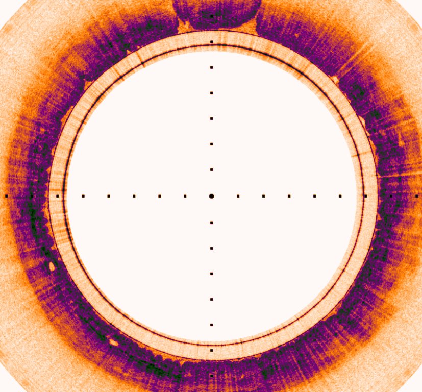
Barrett's Esophagus
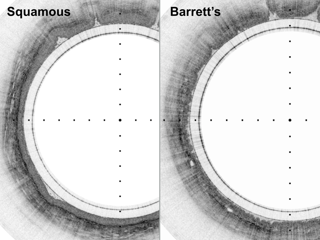
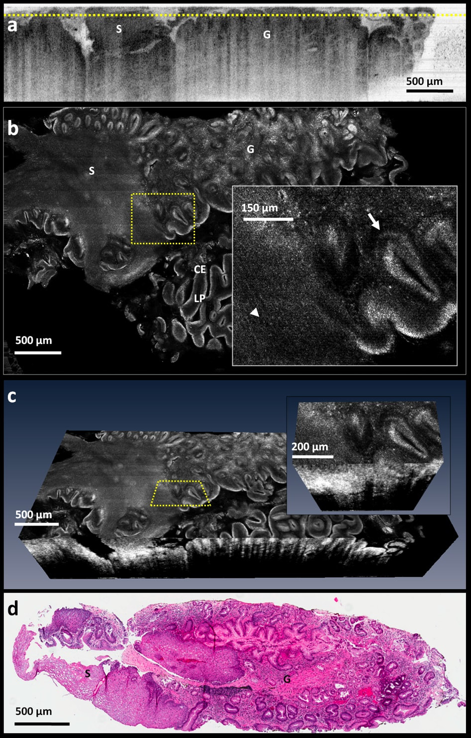
Barrett¹s esophagus (BE) is a condition that is associated with chronic gastroesophageal reflux disease. Early detection of BE is important because it can progress to esophageal adenocarcinoma, a cancer with a high mortality rate. However, when BE is found early it can be monitored to prevent the occurrence of cancer. We have invented a procedure for imaging the entire esophagus with OCT. The images are then used to guide biopsy to evaluate the most severely diseased regions. The Tearney Lab has also developed a tethered capsule that is capable of microscopic imaging a patient¹s GI tract in a simple, low risk, and inexpensive procedure. A patient swallows the capsule and doctors immediately see microscopic images of the digestive tract that are detailed enough to avoid the need for biopsies. This technology is currently being tested in the primary care setting to show the efficacy of capsule screening for Barrett's.

Publications
Gora MJ, Simmons LH, Quénéhervé L, Grant CN, Carruth RW, Lu W, Tiernan A, Dong J, Walker-Corkery B, Soomro A, Rosenberg M, Metlay JP, Tearney GJ. Tethered capsule endomicroscopy: from bench to bedside at a primary care practice. J Biomed Opt. 2016 Oct 1;21(10):104001. doi: 10.1117/1.JBO.21.10.104001. PubMed PMID: 27689919; PubMed Central PMCID: PMC5043371.
Suter MJ, Gora MJ, Lauwers GY, Arnason T, Sauk J, Gallagher KA, Kava L, Tan KM, Soomro AR, Gallagher TP, Gardecki JA, Bouma BE, Rosenberg M, Nishioka NS, Tearney GJ. Esophageal-guided biopsy with volumetric laser endomicroscopy and laser cautery marking: a pilot clinical study. Gastrointest Endosc. 2014 Jun;79(6):886-96. doi: 10.1016/j.gie.2013.11.016. Epub 2014 Jan 23. PubMed PMID: 24462171.
Gora MJ, Sauk JS, Carruth RW, Lu W, Carlton DT, Soomro A, Rosenberg M, Nishioka NS, Tearney GJ. Imaging the upper gastrointestinal tract in unsedated patients using tethered capsule endomicroscopy. Gastroenterology. 2013 Oct;145(4):723-5. doi: 10.1053/j.gastro.2013.07.053. Epub 2013 Aug 8. PubMed PMID: 23932950; PubMed Central PMCID: PMC3866798.
Suter MJ, Vakoc BJ, Yachimski PS, Shishkov M, Lauwers GY, Mino-Kenudson M, Bouma BE, Nishioka NS, Tearney GJ. Comprehensive microscopy of the esophagus in human patients with optical frequency domain imaging. Gastrointest Endosc. 2008 Oct;68(4):745-53. doi: 10.1016/j.gie.2008.05.014. PubMed PMID: 18926183; PubMed Central PMCID: PMC2715833.
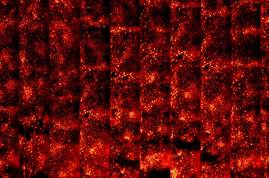
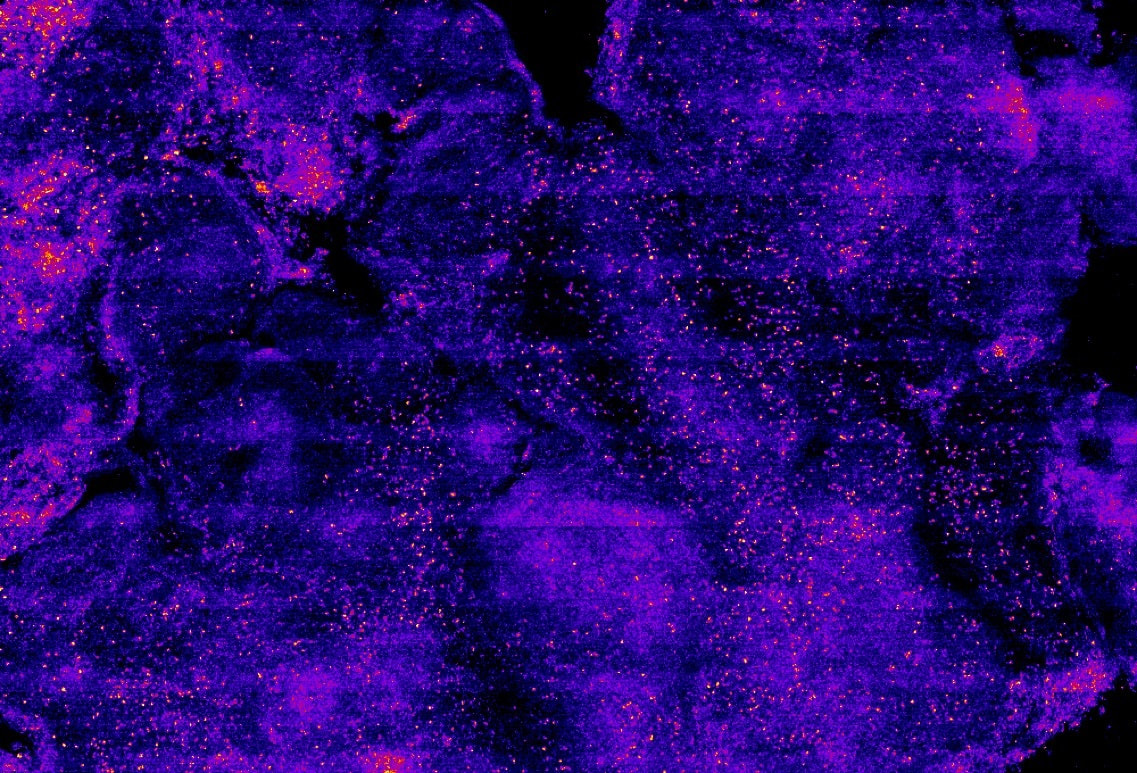
Cystic Fibrosis
The Tearney Lab’s Micro Optical Coherence Tomography (μOCT) technology is being used to help researchers and clinicians better understand and treat cystic fibrosis (CF). CF is one of the most common lethal genetic disorders in the United States. With cystic fibrosis the basic mucus clearance mechanism of the lungs does not work well. The inability to clear the lungs of mucus causes chronic lung infections that may lead to an early death. Researchers and clinicians still do not know exactly why the mucus clearance mechanism of the lungs fails in CF patients. μOCT can directly image this mechanism in action, including the microscopic cilia responsible for driving mucus flow, helping to answer previously unanswerable questions about cystic fibrosis and evaluate treatment methods meant to restore mucus clearance in CF patients.

Publications
Cui D, Chu KK, Yin B, Ford TN, Hyun C, Leung HM, Gardecki JA, Solomon GM, Birket SE, Liu L, Rowe SM, Tearney GJ. Flexible, high-resolution micro-optical coherence tomography endobronchial probe toward in vivo imaging of cilia. Opt Lett. 2017 Feb 15;42(4):867-870. doi: 10.1364/OL.42.000867. PubMed PMID: 28198885.
Liu L, Shastry S, Byan-Parker S, Houser G, K Chu K, Birket SE, Fernandez CM, Gardecki JA, Grizzle WE, Wilsterman EJ, Sorscher EJ, Rowe SM, Tearney GJ. An autoregulatory mechanism governing mucociliary transport is sensitive to mucus load. Am J Respir Cell Mol Biol. 2014 Oct;51(4):485-93. doi: 10.1165/rcmb.2013-0499MA. PubMed PMID: 24937762; PubMed Central PMCID: PMC4189485.
Liu L, Chu KK, Houser GH, Diephuis BJ, Li Y, Wilsterman EJ, Shastry S, Dierksen G, Birket SE, Mazur M, Byan-Parker S, Grizzle WE, Sorscher EJ, Rowe SM, Tearney GJ. Method for quantitative study of airway functional microanatomy using micro-optical coherence tomography. PLoS One. 2013;8(1):e54473. doi: 10.1371/journal.pone.0054473. Epub 2013 Jan 23. PubMed PMID: 23372732; PubMed Central PMCID: PMC3553101.
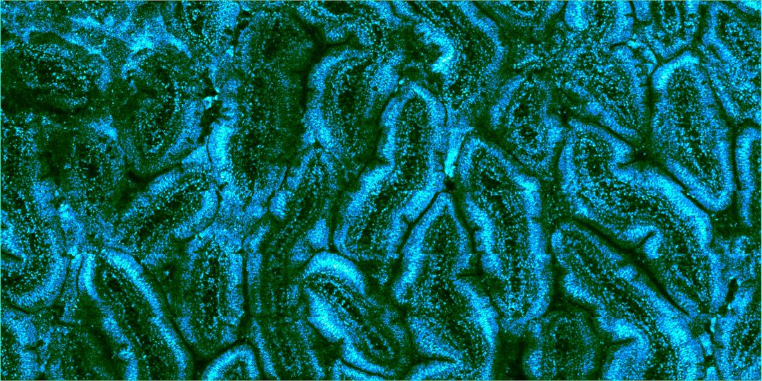
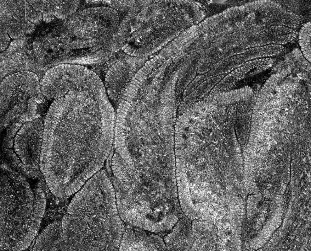
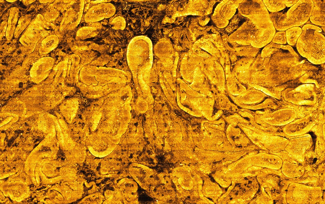
Celiac Disease
Celiac disease is an autoimmune disorder of the small intestine caused by ingestion of gluten that afflicts roughly 1% of the population of the United States. The only known treatment for Celiac disease is a strict gluten-free diet. Traditionally, the diagnosis of celiac disease necessitates endoscopic biopsy, which has a high rate of false negative results. Less invasive SECM and high-resolution OCT tethered capsule microscopes have the potential to diagnose the disease more accurately.